Home
Uncategories
Human Bone Anatomy Diagram / Human Skeleton Worksheet Teachers Pay Teachers / The free science images and photos are perfect learning tools, great for adding to science projects and provide lots of interesting information you may have not known about the human body.
Human Bone Anatomy Diagram / Human Skeleton Worksheet Teachers Pay Teachers / The free science images and photos are perfect learning tools, great for adding to science projects and provide lots of interesting information you may have not known about the human body.
Human Bone Anatomy Diagram / Human Skeleton Worksheet Teachers Pay Teachers / The free science images and photos are perfect learning tools, great for adding to science projects and provide lots of interesting information you may have not known about the human body.. When the arm is spun so that the thumb point to the outside of the body, meaning the palm of the hand looks forward then it is said the hand is supinated.but when the thumb remains in the inside and the palm. Learn more about human anatomy with these free resources. Human organs & anatomy diagram picture category: There are numerous types and combinations of these worksheets, and they can be found in virtually every medical classroom, no matter size or age the students. Human back muscles and bones 12 photos of the human back muscles and bones human back muscles and bones, bone, human back muscles and bones.
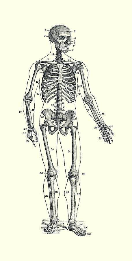
Find free pictures, photos, diagrams, images and information related to the human body right here at science kids. The axial skeleton and the appendicular skeleton. The human skeleton anatomy chart shows three views of the human skeleton (front, back and side) and is painstakingly labeled and painted, producing one of the most captivating and beautiful anatomical charts available. Learn vocabulary, terms, and more with flashcards, games, and other study tools. Start studying human skeleton anatomy.
Forward Facing Skeletal Diagram Vintage Anatomy Poster 2 Drawing By Vintage Anatomy Prints from images.fineartamerica.com Altogether, the skeleton makes up about 20 percent of a person's body weight. This diagram depicts human skeleton with parts and labels. Human back muscles and bones 12 photos of the human back muscles and bones human back muscles and bones, bone, human back muscles and bones. This bodytomy post has detailed information on the human skeletal system, along with a simple diagram, which will help you learn and understand human anatomy. Vertebrae, bones, joints, ligaments, muscles, muscular system, fascia, arteries, veins, nerves and various adjacent organs. It is composed of 300 bones at birth, but later decreases to 80 bones in the axial skeleton and 126 bones in the appendicular skeleton. The pubis, ischium, and ilium together constitute the pelvis while the thigh bone is the femur. Related posts of bones of the hand diagram human back muscles and bones.
Front view of muscles , back view of muscles , organs , nervous system
Using this atlas of human anatomy of the spine and back. The human skeleton anatomy chart shows three views of the human skeleton (front, back and side) and is painstakingly labeled and painted, producing one of the most captivating and beautiful anatomical charts available. We are pleased to provide you with the picture named human skeleton bone diagram.we hope this picture human skeleton bone diagram can help you study and research. Check out pictures and diagram related to bones, organs, senses, muscles and much more. Find diagram of human body organs now. The human skeletal system consists of all of the bones, cartilage, tendons, and ligaments in the body. A diagram of the human skeleton showing bone and cartilage. The structure of a long bone allows for the best visualization of all of the parts of a bone ().a long bone has two parts: There are numerous types and combinations of these worksheets, and they can be found in virtually every medical classroom, no matter size or age the students. Learn more about human anatomy with these free resources. Learn vocabulary, terms, and more with flashcards, games, and other study tools. At your doorstep faster than ever. In addition, the broad hip bones provide protection to the delicate internal organs of the pelvis, such as the intestines, urinary bladder, and uterus.
The femur is a type of long bone located in the thigh and is the largest bone of the skeletal system. Using this atlas of human anatomy of the spine and back. Left hand posterior (dorsal) view the wrist is a complex joint that bridges the hand to the forearm. Altogether, the skeleton makes up about 20 percent of a person's body weight. A diagram of the human skeleton showing bone and cartilage.
Ambesonne Human Anatomy Diagram Of Human Skeleton System With Titled Main Parts Of Body Joints Picture Single Shower Curtain Wayfair from secure.img1-fg.wfcdn.com There are numerous types and combinations of these worksheets, and they can be found in virtually every medical classroom, no matter size or age the students. Its lower end helps create the knee joint. The humerus is the (upper) arm bone. Search for diagram of human body organs now. Posted in diagrams | tagged all bones, human skeleton, skelet, skeleton human eye featured. Left hand posterior (dorsal) view the wrist is a complex joint that bridges the hand to the forearm. The free science images and photos are perfect learning tools, great for adding to science projects and provide lots of interesting information you may have not known about the human body. The human skeleton anatomy chart shows three views of the human skeleton (front, back and side) and is painstakingly labeled and painted, producing one of the most captivating and beautiful anatomical charts available.
674 x 599 photo description:
The humerus is the (upper) arm bone. The hip itself is a ball and socket joint, much like the shoulder.the structures necessary to create this joint are the socket, the joint capsule, muscle, ligaments, and the neck. Search for diagram of human body organs now. It joins with the scapula above at the shoulder joint (or glenohumeral joint) and with the ulna and radius below at the elbow joint. These bones are arranged into two major divisions: In addition, the broad hip bones provide protection to the delicate internal organs of the pelvis, such as the intestines, urinary bladder, and uterus. Human skeleton bones labeled 12 photos of the human skeleton bones. Free shipping on all orders over $35. For more anatomy content please follow us and visit our website: Posted on may 28, 2014 by admin. The axial skeleton runs along the body's midline axis and is made up of 80 bones in the following regions: For teachers, students, health professionals, or anyone interested in learning about the anatomy of the human body. The femur and/or hip may fracture secondary to trauma, so understanding the femur bone anatomy is important.
There are numerous types and combinations of these worksheets, and they can be found in virtually every medical classroom, no matter size or age the students. For more anatomy content please follow us and visit our website: In addition, the broad hip bones provide protection to the delicate internal organs of the pelvis, such as the intestines, urinary bladder, and uterus. A diagram of the human skeleton showing bone and cartilage. The structure of a long bone allows for the best visualization of all of the parts of a bone ().a long bone has two parts:
1 from Check out pictures and diagram related to bones, organs, senses, muscles and much more. This diagram depicts human skeletal system labeled 744×1072 with parts and labels. The femur and/or hip may fracture secondary to trauma, so understanding the femur bone anatomy is important. The femur, or thighbone, is the longest and largest bone in the human body. The diaphysis and the epiphysis.the diaphysis is the tubular shaft that runs between the proximal and distal ends of the bone. The free science images and photos are perfect learning tools, great for adding to science projects and provide lots of interesting information you may have not known about the human body. The humerus is the (upper) arm bone. Free shipping on all orders over $35.
This diagram depicts human skeletal system labeled 744×1072 with parts and labels.
Check out pictures and diagram related to bones, organs, senses, muscles and much more. For more anatomy content please follow us and visit our website: The anatomy of the femur can be divided into proximal, central, distal, and posterior parts. The hip itself is a ball and socket joint, much like the shoulder.the structures necessary to create this joint are the socket, the joint capsule, muscle, ligaments, and the neck. The humerus is the (upper) arm bone. The axial skeleton runs along the body's midline axis and is made up of 80 bones in the following regions: When the arm is spun so that the thumb point to the outside of the body, meaning the palm of the hand looks forward then it is said the hand is supinated.but when the thumb remains in the inside and the palm. Learn vocabulary, terms, and more with flashcards, games, and other study tools. These bones are arranged into two major divisions: Start studying human skeleton anatomy. Front view of muscles , back view of muscles , organs , nervous system Human organs & anatomy diagram picture category: For teachers, students, health professionals, or anyone interested in learning about the anatomy of the human body.
The human skeleton anatomy chart shows three views of the human skeleton (front, back and side) and is painstakingly labeled and painted, producing one of the most captivating and beautiful anatomical charts available human bone anatomy. The diaphysis and the epiphysis.the diaphysis is the tubular shaft that runs between the proximal and distal ends of the bone.

0 Comments:
Posting Komentar